Neurological autoimmune diseases
1. Neuropathies
Myelin Associated Glycoproteins (MAG)
Peripheral neuropathies, autoimmune reactions of the peripheral nervous system, are associated with autoantibodies to various neural glycoconjugates.
Neuropathies associated with anti-MAG with IgM paraproteinemia often progress slowly, with evidence of demyelination and a variable degree of axonal loss associated with ataxia.
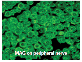
Immunofluorescence is a sensitive method for the screening and detection of anti-myelin-associated proteins and ganglioside autoantibodies.
2. Paraneoplastic Syndrome
Neuronal Antibodies
Autoimmune reactions of the central nervous system, which are recognized as paraneoplastic neurological disorders, are manifestations of an anti-tumor immune response.
The following autoantibodies occur in paraneoplastic syndromes:
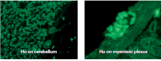
- Anti-Hu, Type I Antineuronal Core Antibodies (ANNA-1):
- is associated with small cell lung cancer leading to paraneoplastic encephalomyelitis (PE)
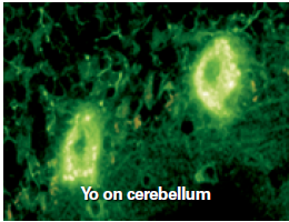
- Anti-Yo Antibodies to Purkinje Cells (PCA-1):
- are associated with ovarian and breast carcinomas leading to paraneoplastic cerebellar degeneration (PCD)
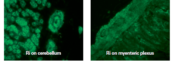
- Anti-Ri type II Anti-Neuronal Core Antibodies (ANNA-2):
- is associated with neuroblastoma (children) and fallopian or breast cancers (adults) resulting in Paraneoplasticopsoclonus myoclonus ataxia (POMA)
These markers help to distinguish between true paraneoplastic disorders and other inflammatory disorders of the nervous system that mimic a paraneoplastic syndrome.

| Hu and Ri autoantibodies stain the granular cell nucleus and are easily distinguishable from the Purkinjee cell cytoplasm-staining Yo antibodies! |
3. Myasthenia Gravis (MG)
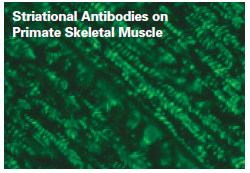
Striational Muscle Antibodies
Myasthenia gravis is caused by a number of associated autoantibodies.
These include antibodies to skeletal muscle detected by immunofluorescence using primate skeletal muscle tissue.
Significant titers of striation antibodies occur in myasthenia gravis mainly in association with thymomas. A positive strip antibody with negative results for the acetylcholine receptor antibody may support the diagnosis of acquired MG and may indicate a thymoma.
Determination of Antibodies in Neurological Autoimmune Diseases
Determination of Myelin Associated Glycoproteins (MAG) and Neuronal Antibodies
- IF neural antibodies and MAG antibodies
- Western Blot Neuronal Antibodies and MAG Antibodies
Determination of Myasthenia Gravis Antibody
- Primates Skeletal Muscle
- Rats Skeletal Muscle

 Deutsch


