In recent years, three-dimensional (3D) cell culture models have gained popularity over two-dimensional (2D) models for developing new drugs and are also increasingly used in fundamental research. Due to their three-dimensionality, they simulate properties of designated organs better than 2D models and show more relevant results in comparison. Especially in the last five years a rapid development of 3D models was noticeable. This led to a trend in research concluding that 3D models could even be used to simulate in-vivo conditions.
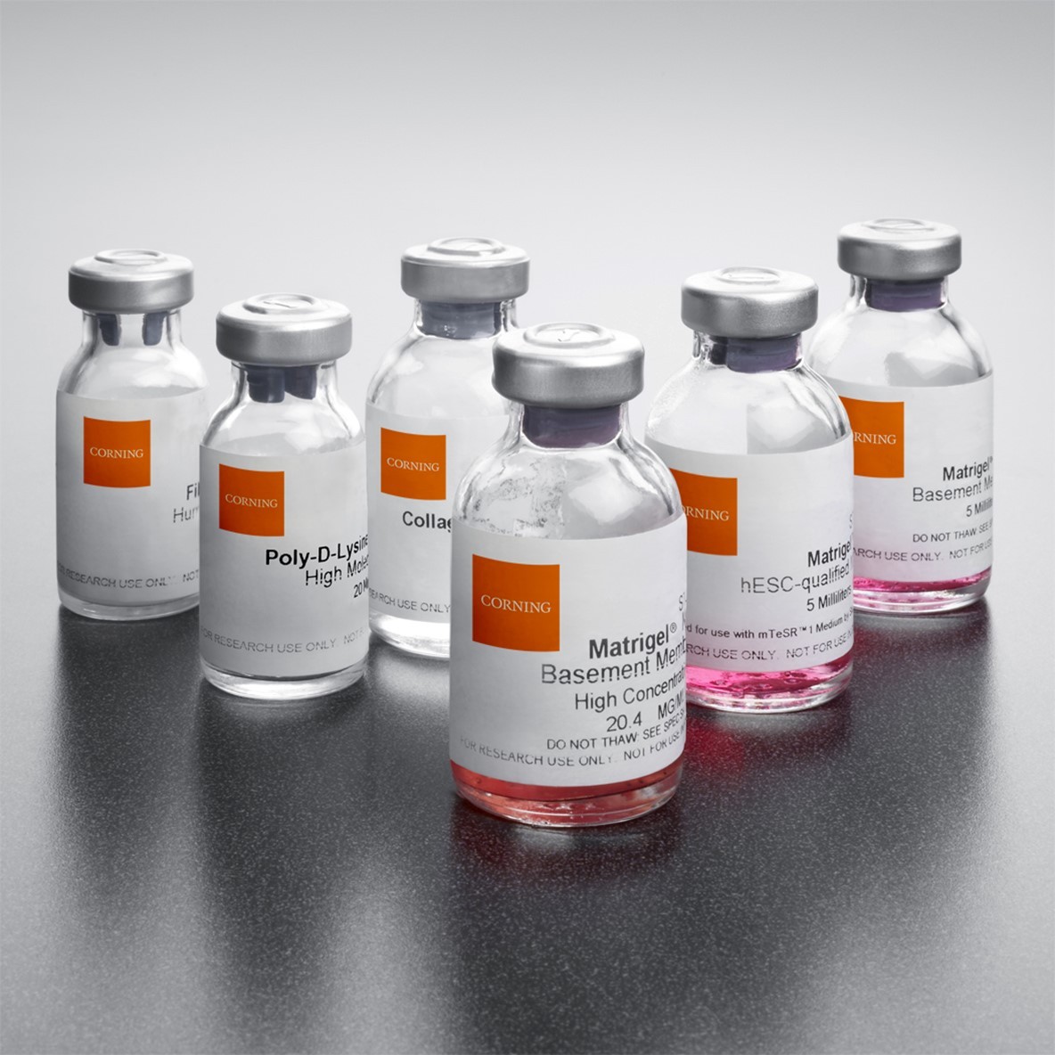
 Most in-vitro tests are still based on 2D cell models cultivated on treated glass or plastic surfaces. However, they fail to take tissue-specific properties or the microenvironment, caused by interacting with other cells, into account. Especially for fundamental and translational research (e.g. in oncology), cell models capable of simulating interactions with other cell types are needed. For this purpose, 3D cell culture models with the ability to display in-vivo like cell functions have been increasingly developed in recent years. In contrast to conventional 2D models, 3D models consider the three-dimensionality of the tissue. They are either cultivated on structural proteins such as collagen, gelatin-methacrylate or Matrigel or are dissected from tissue sections. Milestones in the development of 3D cell culture models have been reached with organoids and spheroids, 3D printing applications as well as oncological models for high-throughput screening. With these models, it is possible to investigate the efficacy of therapeutics while observing cell metabolism and cell growth simultaneously. Analysis tools for this purpose were significantly improved as well.
3D-tumor spheroid models
Most cell lines form 3D spheroid with a diameter of 30-50 µm, but some have the ability to grow to a larger size. Significant for spheroids are the ability to display a natural metabolism and tumor-like growth.
3D-bioprinting application
A very new field is represented by 3D bioprinting. Only this year researchers from the University of Innsbruck successfully printed a live 3D skin model which formed vascular cells spontaneously. This was done by printing living cells on bioactive protein gels on a chip. Within six to eight days a three-layered skin model was formed which had the ability to build connective tissue and grow blood vessels on its own as well as differentiating an epidermis layer.
Organoid models on chips
One of the most promising applications for 3D cell culture technologies is the usage of organoid models in which stem cells are used to differentiate into tissue samples of different organs. These can be connected on a chip with a fluidic system, mimicking live metabolism.
Advances in research
The development of organoids on chips has led to new possibilities and enabled, for instance, investigation of electrical activity of heart cells in real time. Additionally, the development of personalized therapeutic strategies using stem cells is within reach: medical staff are not only able to grow cells or tissues for transplants but are also able to test medication for individual side-effects prior to subscribing them to patients.
Outlook – market growth for 3D cell culture models
The versatile applications of 3D models have led to a two-digit growth rate in recent years. However, the maximum capacity of these models has not yet been reached. With the rapid development of 3D cell culture models simulating in-vivo conditions, they have the potential to replace animal studies for medical research in the long run.
3D-cell culture models at SZABO-SCANDIC
Here at Szabo-Scandic, we provide the best products from Corning and Lonza for 3D cell culture and have a suitable model for any research question or requirements.

 Deutsch

